We often hear the terms sonogram and ultrasound used interchangeably. But are they the same thing?
What is the Difference Between the Two?
There is a slight difference in the terms but, simply, one is the process and the other is the product. An ultrasound is a procedure that uses sound waves to create images of organs and soft tissues. The sonogram is the actual image that is created by the ultrasound. So the difference is a matter of semantics.
How Does an Ultrasound Work?
Ultrasounds use high-frequency sound waves in the analysis and diagnosis of medical conditions. These sound waves are transmitted and echo off the tissues and organs that are being studied. They can only create images when there is a surface to reflect from. When they hit a surface, they reflect or “echo” back and create images. These images enable your doctor to study what is going on inside the body. Ultrasounds are noninvasive, safe and require no radioactive components.
While most of us are familiar with ultrasounds that are used in obstetrics and pregnancy to monitor a baby’s health, there are many other uses for sonograms:
- Gallbladder disease diagnosis
- Blood flow evaluation
- Breast lump examination
- Guidance for a biopsy
- Thyroid evaluation
- Genital and prostate issues
- Joint inflammation assessment
- Metabolic bone disease evaluation
Other variations of ultrasounds are also used for many different types of diagnostics:
- Doppler ultrasounds are used to measure blood circulation in blood vessels and in the heart to determine abnormal blood flow.
- Elastography can be used to determine the elastic properties or stiffness of tissues to make a distinction of diseased tissue and tumors from healthy tissue.
- Bone sonography is used to determine bone density.
- Therapeutic ultrasounds are used in physical therapy to soften and break up tissues in the body including muscles, joints and ligaments.
While most ultrasounds are performed on the skin’s surface using what is called a transducer, other ultrasounds get a more detailed image by inserting a specialized transducer into a body cavity. Some of these include
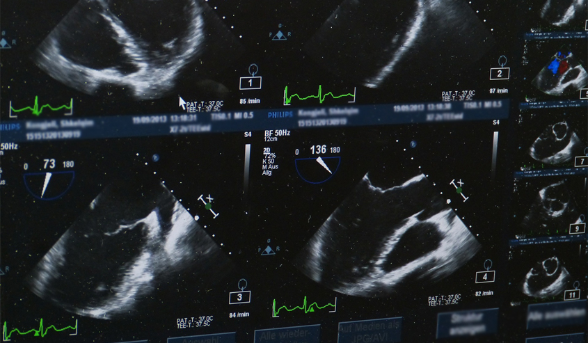
- Transesophageal echocardiogram utilizes a specialized probe inserted into the esophagus to get images of the heart.
- Transvaginal ultrasound uses a specialized transducer wand to get images of the uterus and ovaries.
- Transrectal ultrasound uses a transducer wand in the rectum to diagnose prostate conditions.
Although ultrasounds and sonograms are extremely helpful for evaluating and understanding many conditions, they do have limitations. Because sound waves don’t transmit well through dense bone or through air, an ultrasound is not as effective in imaging certain body parts such as the lungs or head. Doctors will use different imaging techniques for these areas of the body.
At Vital Imaging, we offer ultrasounds and many other imaging services. Our team of caring professionals is there for you at all times, making sure you are comfortable and answering any of your questions. To learn more about our services or to schedule an appointment, call (305) 596-9992.