
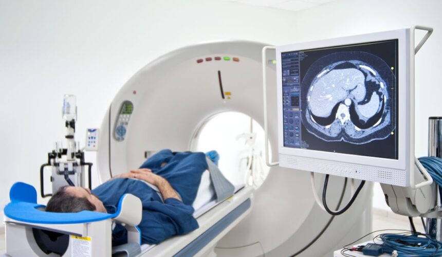
A CT scan for kidney stones is quick, painless, and usually done without contrast dye. For most Miami patients with sudden side or back pain, it is the test doctors reach for first. The scan does more than say “yes, there’s a stone.” It measures the stone and shows exactly where it sits. Those two facts decide whether you wait it out or see a urologist. Why Doctors Order a CT Scan for Kidney Stones Doctors order a CT because it finds stones better than any other test. A low-dose CT without dye catches about 97% of stones, per the...
Blog

Low-Dose CT Lung Cancer Screening in Miami: Who Qualifies
You likely qualify for low-dose CT lung cancer screening if you are 50 to 80 years old, have a 20 pack-year smoking history, and still smoke or quit within the past 15 years. Those are the national rules from the U.S. Preventive Services Task Force. Medicare uses a narrower age range, and that gap surprises many people. The scan takes minutes. The eligibility details are where most Miami patients get stuck. Who Qualifies for Low-Dose CT Lung Cancer Screening in Miami? You qualify if you meet all three rules at the same time. Miss one, and the scan no longer...
Spine MRI in Miami: Cervical, Thoracic, and Lumbar Explained
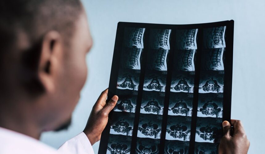
A spine MRI in Miami is really three separate exams: cervical for the neck, thoracic for the mid-back, and lumbar for the low back. Your doctor chooses the section based on where your symptoms start, which isn’t always where they hurt most. Each section gets its own time slot, its own set of images, and often its own insurance approval. Which Part of the Spine Does Each MRI Cover? The cervical spine is the 7 bones in your neck. The thoracic spine is the 12 bones in your mid-back, where the ribs attach. The lumbar spine is the 5 bones...

Pre-Surgical Diagnostic Clearance Testing in Miami
Pre-surgical diagnostic testing in Miami usually means a short list of heart and chest tests that show whether your body can safely handle anesthesia and surgery. The list varies by patient. It depends on your health, age, and the size of the operation. The biggest problem is rarely the test itself. It’s usually results that arrive late or tests that expire before your surgery date. Is Clearance Testing the Same as Surgical Planning Imaging? No. Clearance testing asks whether you can safely go through surgery. Planning imaging shows the surgeon exactly what to fix. Mixing them up causes last-minute delays....

How Diagnostic Testing Works for Workers’ Comp Claims in Miami
Workers’ comp diagnostic testing in Miami runs on carrier authorization, not on your doctor’s judgment alone. Under Florida law, the insurance carrier selects your treating physician, and that physician cannot send you to an imaging center without the carrier’s prior approval. The scan itself is the easy part. Getting the paperwork lined up determines whether you wait three days or three weeks. Who Decides Where You Get Your Scan The carrier does. Florida’s workers’ compensation medical rules give the employer and its insurance carrier the right to select your authorized treating physician, and that physician’s referrals route through the carrier...

Family History of Breast Cancer: When to Start Screening Early
If breast cancer runs in your family, your screening should usually start before 40. A mammogram family history plan in Miami is built on two numbers: how old your relative was at diagnosis, and what your calculated lifetime risk turns out to be. The American College of Radiology recommends every woman get a formal risk assessment by age 25 so that math happens on time. Most women never get one, and that is where early screening quietly gets lost. Which Relatives Actually Count First-degree relatives carry the most weight: mother, sister, daughter. Second-degree relatives such as grandmothers, aunts, and half-sisters...
How the BI-RADS Mammogram Scoring System Works
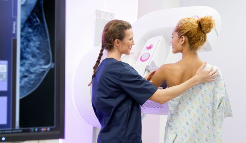
BI-RADS is a number from 0 to 6 that a radiologist assigns to your mammogram to say what was found and what happens next. Every accredited BI-RADS mammogram center in Miami uses the same seven categories, so a 3 at one facility means the same thing as a 3 at another facility. The number is not a cancer score. It is a management instruction. Once you know how to read it, the rest of the report gets much easier. What the Seven BI-RADS Categories Mean Each category pairs a finding with an action. Here is the full scale in plain...

Walk-In Diagnostic Testing in Miami: No Appointment Needed
Some diagnostic tests in Miami really are walk-in. Others only look that way. At a walk-in diagnostic center in Miami, what decides your visit is rarely the appointment book. It is whether you arrive holding a valid order from a treating provider. Get that part right and a same-day X-ray can take less time than a lunch break. What “Walk-In” Really Means at an Imaging Center Walk-in means we can fit you into an open slot the same day. It does not mean we can scan you without an order. That distinction trips up more Miami patients than anything else,...
Bilingual Mammogram Centers in Miami: Why It Matters for Care Quality
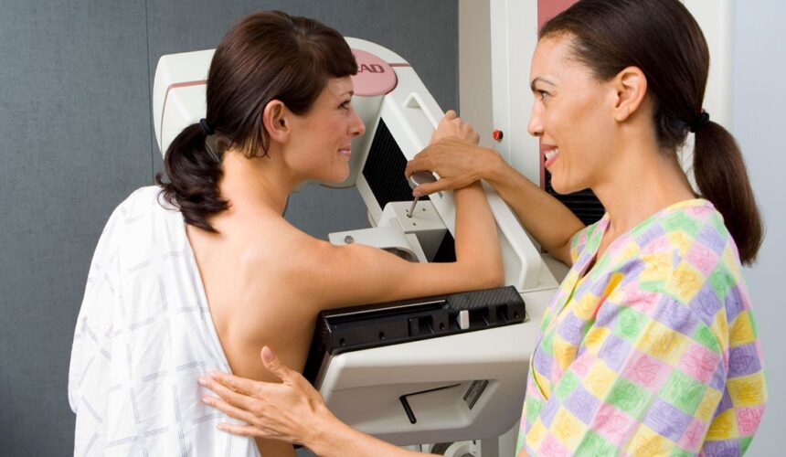
A bilingual mammogram center changes the quality of your care, because nearly everything that makes the exam useful depends on communication. You describe symptoms, answer history questions, follow positioning instructions, and later read a written report. Anything lost in translation lowers what your radiologist can see and what you can act on. In a county where roughly three out of four residents speak a language other than English at home, that gap is common. What Language Access Really Looks Like at an Imaging Center Real language access covers five touchpoints, not one. A greeting in Spanish at the front desk...
Walk-In vs. Scheduled Mammograms in Miami: Pros and Cons
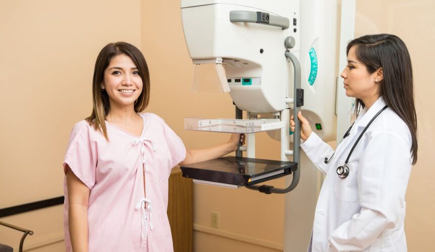
A walk-in mammogram is a fine choice for a routine yearly screening, and a scheduled appointment is the safer choice for everything else. The split comes down to one question: do you have symptoms or a reason your doctor wants a closer look? Convenience is real, and so is the risk of showing up on the wrong day for the wrong exam. Here is how the 2 options compare in practice. What a Walk-In Mammogram Actually Includes Walk-in service almost always means a screening mammogram and nothing more. A screening exam is a routine check for women with no symptoms....
Open vs. Closed MRI in Miami: Which One Is Right for You?
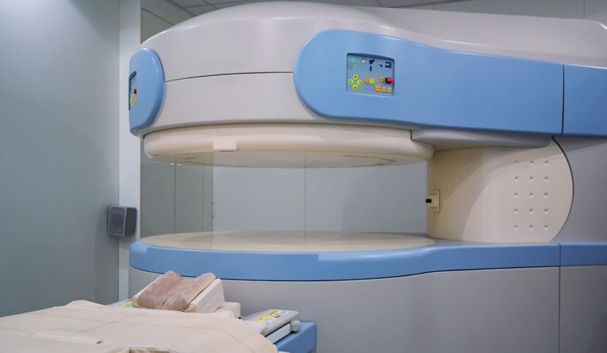
For most scans, a closed MRI is the better choice because its stronger magnet produces sharper images and your doctor gets a clearer answer. The open vs. closed MRI question really matters when you cannot fit inside a standard machine or cannot stay calm in a tight space. Those are real problems, and an open scanner solves them. Before you pick a machine based on comfort alone, it helps to know what you trade away. How Open and Closed MRI Machines Are Built Differently A closed MRI is a tube. You slide into a cylinder that is open at both...
How to Choose an MRI Center in Miami: 8 Questions to Ask Before Booking
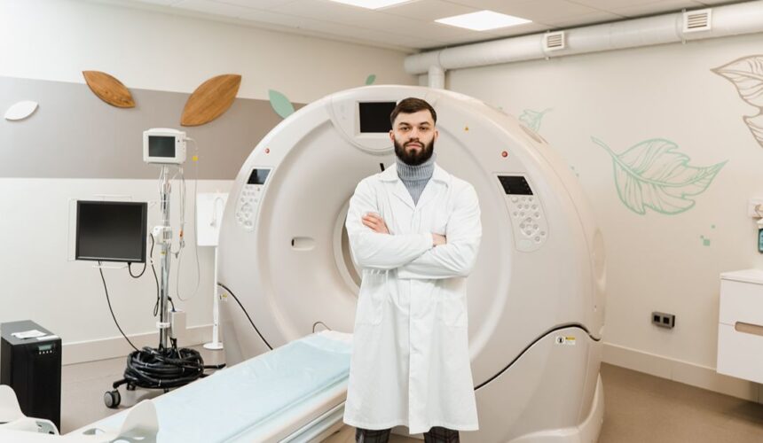
The best MRI center in Miami is the one with current accreditation, a scanner that fits the body part your doctor ordered, and a radiologist who reads that type of study every day. Cost and wait time are very important, but they come second. Most people book the first place their doctor’s office sends the order to, and never ask a single question. 10 minutes on the phone can save you a repeat scan, a surprise bill, or a month of waiting. What Sets One MRI Center in Miami Apart From Another Two centers can charge the same price and...
Cyst vs Tumor: Main Differences
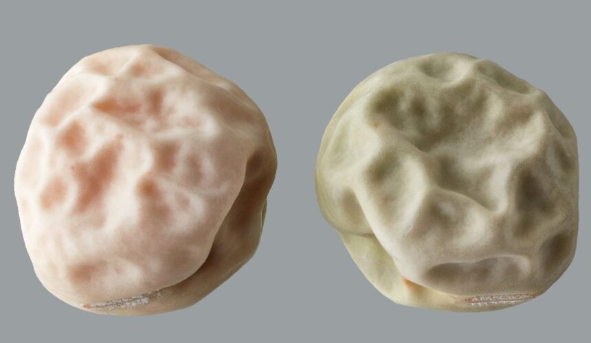
The difference between a cyst and a tumor is simple: a cyst is usually a fluid-filled sac, while a tumor is a solid mass of tissue. Knowing the differences helps you understand lumps and when to seek care. What’s the Difference Between a Tumor and a Cyst? The main difference between a tumor and a cyst is their composition. A cyst is filled with fluid, air, or soft material. A tumor is made of solid tissue. Cysts are often harmless, while tumors can be benign or cancerous. Quick comparison Cyst: Fluid-filled, usually benign Tumor: Solid mass, can be benign or...

Dense Breast Tissue: Why You Must Know What It Means
Dense breast tissue means you have more gland and connective tissue than fat in your breasts. This matters because it can make it harder to find cancer on imaging tests. What Is Dense Breast Tissue? Dense breast tissue is a common condition in which the breast contains more fibrous and glandular tissue and less fat. It is not a disease, but it affects how scans look. Dense tissue looks white on imaging. Cancer also looks white. This makes it harder to tell them apart. Key facts Many women have dense breasts It is normal and common It does not mean...
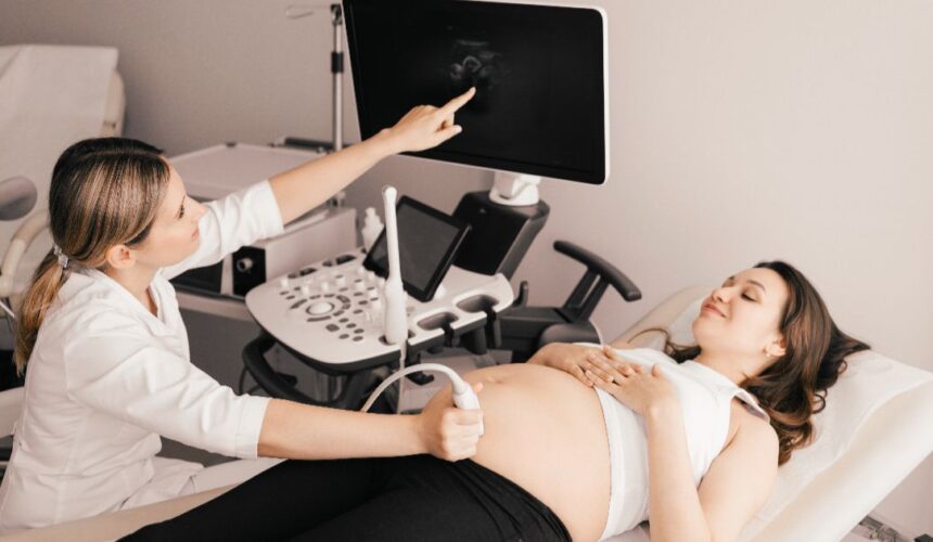
Gynecological Ultrasounds: Everything You Need to Know
Gynecological ultrasounds are imaging tests that use sound waves to create pictures of the female reproductive system. It is used to check organs, find problems early, and monitor pregnancy. What Are Gynecological Ultrasounds? Gynecological ultrasounds use sound waves to create images of organs like the uterus, ovaries, and breasts. They do not use radiation. The machine sends sound waves into the body. The waves bounce back, creating an image on a screen. Why doctors use them Check for cysts or tumors Monitor pregnancy Find causes of pain or bleeding Track reproductive health This test is safe, quick, and widely used....
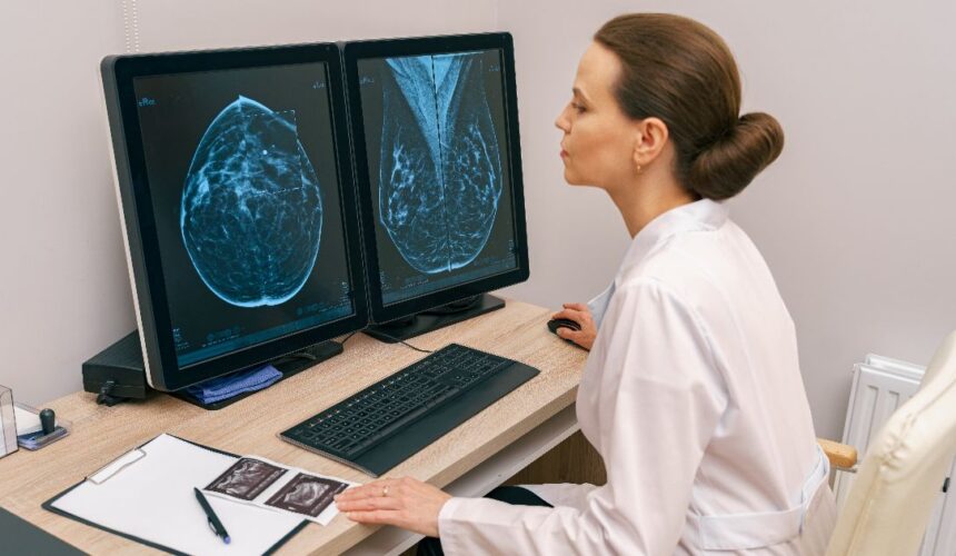
3D Tomosynthesis Mammogram: Complete Guide
A 3D tomosynthesis mammogram is an advanced breast imaging test that creates detailed layered images of the breast. It helps doctors find cancer earlier and with more accuracy than standard mammograms. What Is a 3D Tomosynthesis Mammogram? A 3D tomosynthesis mammogram is a type of breast X-ray that takes many images from different angles. These images are then combined to form a 3D view of the breast. It lets doctors see “slices” of breast tissue rather than a single flat image. This makes it easier to spot small tumors or hidden areas. Importance It reduces the overlap of tissue It...
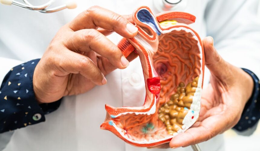
What Is an ERCP (Endoscopic Retrograde Cholangiopancreatography)?
An ERCP is a medical procedure that diagnoses and treats problems in the bile ducts and pancreatic ducts. Endoscopic retrograde cholangiopancreatography (ERCP) uses a flexible scope and X-ray imaging to find and remove blockages. This procedure combines endoscopy and fluoroscopy. Endoscopy uses a thin tube with a camera. Fluoroscopy uses X-ray guidance. Together, they allow doctors to see inside the ducts and treat problems simultaneously. ERCP is both a diagnostic and treatment procedure. What Does an ERCP Do? An ERCP examines and treats blockages in the bile ducts and pancreatic ducts. It can remove stones, open narrow ducts, and place...
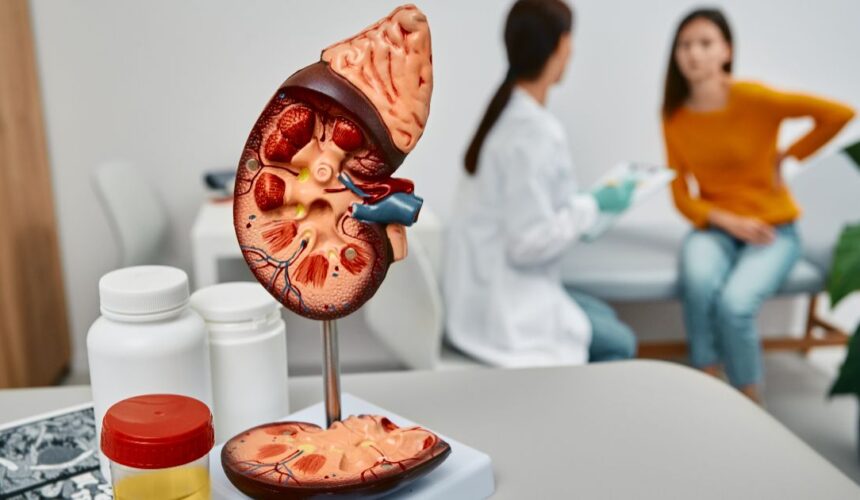
What Is a Blocked Bile Duct?
A blocked bile duct means bile can’t move from your liver to your small intestine. Bile is a fluid your liver makes to help digest fats. It travels through small tubes called bile ducts. When one of these tubes gets blocked, bile builds up in your body. This can hurt your liver and cause infection. Getting diagnosed early is important. Bile Duct Obstruction Bile duct obstruction is a blockage that prevents bile from draining properly. The blockage can happen in the liver, in the common bile duct, or near the pancreas. When bile cannot flow out, it backs up into...