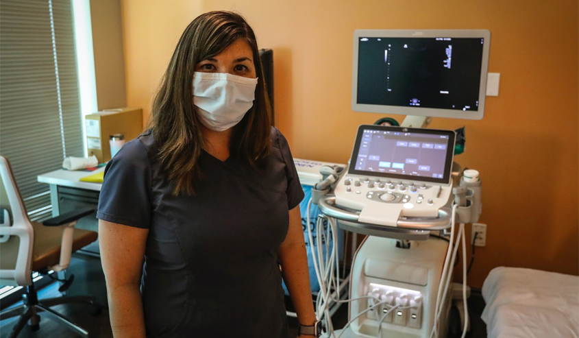
So is it a sonogram or an ultrasound?
Often used interchangeably, one is the process, and the other is the product. An ultrasound is the procedure that uses sound waves to create images of the baby and uterus during pregnancy. The sonogram is the actual image that is created by the ultrasound.
Many of us are familiar with ultrasounds as they apply to pregnancy. They allow a physician to monitor both baby in utero and mother to ensure that all is well during gestation. As an important prenatal test, it utilizes sound waves to create an image of the baby and its position in the womb. At later points, it is able to offer clear images of arms, legs and even facial features. And, yes, even the baby’s gender.
When is an Ultrasound Done?
Ultrasounds will typically be ordered when a woman is in the first and second trimester of her pregnancy. Most physicians will wait until at least 6 weeks to order a first trimester ultrasound but the gestational sac can be seen as early as 4 ½ weeks and the heartbeat as early as 5 to 6 weeks. Ultrasounds are ordered in varying numbers and timing depending on the mother and baby and their unique health situations.
An ultrasound can give the doctor important information such as:
- Pregnancy confirmation
- Check for multiples
- Determine due date
- Check baby’s age and growth
- Check baby’s heartbeat, movement and development
- Check for position in utero prior to delivery
- Examine the mother’s ovaries and uterus
A physician will also use ultrasound technology to screen for the baby’s health and look for birth defects. Ultrasounds are often used to support other testing such as chorionic villus sampling, amniocentesis, biophysical profiles, nuchal translucency screening and to further test for pregnancy complications.
Is an Ultrasound Safe?
Ultrasounds have been used for over 30 years. Because they are non-invasive and use no radiation, they are safe for both mother and baby.
During a transabdominal ultrasound, a gel is used on the mother’s abdomen and a wand that is rubbed over the area will produce images of the baby. An ultrasound will typically last around 30 minutes. The technician will ask the mother to fill her bladder for the earlier procedures.
First trimester ultrasounds will give the physician good information about the fetal heartbeat and the location of the pregnancy as well as an estimate of a due date. A second trimester ultrasound will give the physician information about how the baby is developing and offer peace of mind that things are going well. This is when parents can choose whether they want to know the gender of their baby.
At Vital Imaging, we offer ultrasounds and many other imaging services. Our team of caring professionals is there for you at all times, making sure you are comfortable and answering any of your questions. To learn more about our services or to schedule an appointment, call (305) 596-9992.
