Bone Density

Bone Density
Bone Density Scans, also known as DEXA scans, use a very small dose of ionizing radiation to produce pictures of the inside of the body (usually the lower (or lumbar) spine and hips) to measure bone loss. It is commonly used to diagnose Osteoporosis, to assess an individual’s risk for developing osteoporotic fractures. DEXA is simple, quick and noninvasive. It’s also the most commonly used and the most standard method for diagnosing Osteoporosis.
What to Expect
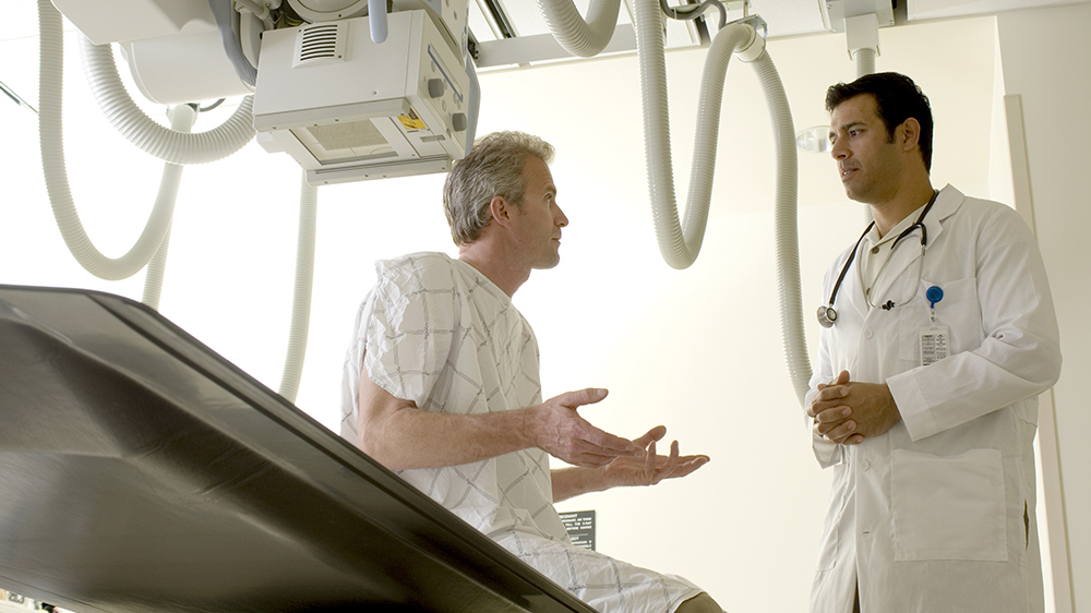
In the central DEXA examination, which measures bone density of the hip and spine, the patient lies on a padded table. An x-ray generator is located below the patient and an imaging device, or detector, is positioned above. The detector is slowly passed over the area of interest, generating images on a computer monitor.
You must hold very still and may be asked to keep from breathing for a few seconds while the x-ray picture is taken to reduce the possibility of a blurred image. The DEXA scan is usually completed within 20-30 minutes.
Preparation
No special preparation required for this exam.
Exams Performed
- Bone Densitometry Scan
- DEXA
- DXA
